Labeled Diagram Of An / Draw A Labelled Diagram Of Spirogyra Cell From Science Diversity In Living Organisms Class 9 Cbse - A neuron is also known as the nerve cell.
Labeled Diagram Of An / Draw A Labelled Diagram Of Spirogyra Cell From Science Diversity In Living Organisms Class 9 Cbse - A neuron is also known as the nerve cell.. Labeled diagram of the human kidney. The majority of the nervous system is tissue made up of two classes of cells: Sepals protect the flowers before they bloom. It is the beginning of the digestive tract and the process of digestion begins from the mouth, where teeth help by breaking and grinding the food. Flowers contain vital parts, including petals, which form flowers.
The majority of the nervous system is tissue made up of two classes of cells: Lungs are an excellent example of how several tissues can be compactly arranged, yet providing a large surface area for gaseous exchange. Diagram of body organs female pics stock illustrations. Labeled diagram of the human kidney. Mitosis is a process of cell division which results in the production of two daughter cells from a single parent cell.
Medial Muscles And Bones Of The Foot Sole Labeled Human Anatomy Diagram Stock Photo Download Image Now Istock from media.istockphoto.com See labeled brain anatomy stock video clips. This diagram depicts labeled diagram of digestive system with parts and labels. Nervous system anatomy nervous tissue. Spend some time studying the diagram to get a clear picture in your mind. We hope this picture labelled diagram of the muscles in the human body can help you study and research. The f 2s is nonbonding. Find a great range of the diagram of human body and anatomy diagrams in the following pictures. It also carriers the microscopic illuminators.
Lungs are an excellent example of how several tissues can be compactly arranged, yet providing a large surface area for gaseous exchange.
Female anatomy includes the external genitals, or the vulva, and the internal reproductive organs. Structure of a motor neuron. This diagram depicts labeled diagram of digestive system with parts and labels. Diagram of parts of a microscope. It also carriers the microscopic illuminators. Who let the hulk out? The majority of the nervous system is tissue made up of two classes of cells: For more anatomy content please follow us and visit our website: The structure of an atom explained with a labeled diagram. Sepals protect the flowers before they bloom. Nervous system anatomy nervous tissue. You should now be able to label all the main anatomical features. It is the beginning of the digestive tract and the process of digestion begins from the mouth, where teeth help by breaking and grinding the food.
Female anatomy includes the external genitals, or the vulva, and the internal reproductive organs. Most flowers have male and female parts that allow the flower to produce seeds. Human eye anatomy, retina, optic disc artery and vein etc. Zoological graphic with birds bones, digestive system and inside structure. National eye institute , national eye health education program subject:
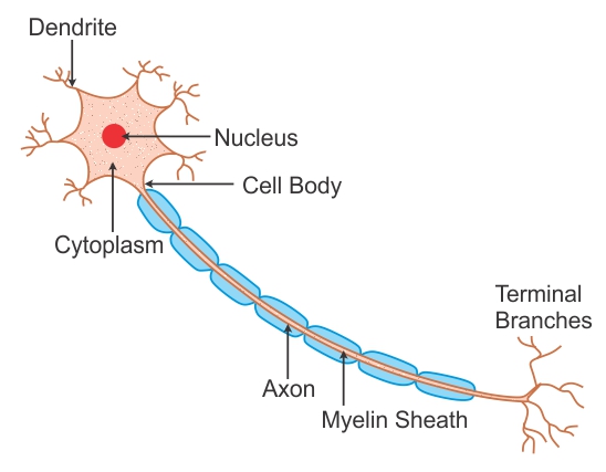
Labeled Diagram Of Nerve Cell Biology Topperlearning Com Wwudaicc from images.topperlearning.com Mo diagram for hf the ao energies suggest that the 1s orbital of hydrogen interacts mostly with a 2p orbital of fluorine. It also carriers the microscopic illuminators. Eye anatomy, eye diagram, cornea, iris, lens, macula, optic nerve, pupil, retina, vitrous gel, diabetic eye disease. A neuron is a specialized cell, primarily involved in transmitting information through electrical and chemical signals. How many of these structures do you recognize from the previous sections? Diagram of parts of a microscope. Let learn the different parts of the human digestive system. Diagram of body organs female pics stock illustrations.
The heart pumps blood through the network of arteries and.
Mouth — it includes teeth, salivary glands and tongue. Labeled biological inner organs scheme chicken anatomy vector illustration. Nervous system anatomy nervous tissue. This diagram depicts labeled diagram of digestive system with parts and labels. The axial skeleton and the appendicular skeleton. The knee is a complex joint that flexes, extends, and twists slightly from side to side. A neuron is a specialized cell, primarily involved in transmitting information through electrical and chemical signals. Find a great range of the diagram of human body and anatomy diagrams in the following pictures. The f 2s is nonbonding. The diagram below shows the structure and functions of the human digestive system. Flowers contain vital parts, including petals, which form flowers. This article looks at female body parts and their functions, and it provides an interactive diagram. Learn more about the main parts of a flower.
Structure of a motor neuron. Who let the hulk out? Nervous system anatomy nervous tissue. A neuron is a specialized cell, primarily involved in transmitting information through electrical and chemical signals. Anatomynote.com found labelled diagram of the muscles in the human body from plenty of anatomical pictures on the internet.
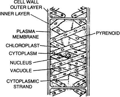
Draw A Labelled Diagram Of Spirogyra Cell From Science Diversity In Living Organisms Class 9 Cbse from www.zigya.com Find a great range of the diagram of human body and anatomy diagrams in the following pictures. For more anatomy content please follow us and visit our website: Labeled biological inner organs scheme chicken anatomy vector illustration. See labeled brain anatomy stock video clips. Plant cell diagram plant cell labeled, 3d plant cell. Posted on october 24, 2015 by admin. Label fractions on number line. They are found in the brain, spinal cord and the peripheral nerves.
Diabetes and healthy eyes toolkit and website keywords:
The heart pumps blood through the network of arteries and. Most seeds transform into fruits and vegetables. Picture), the vegetative phase in which the spores are produced, and what is. Plant cell diagram plant cell labeled, 3d plant cell. We hope this picture labelled diagram of the muscles in the human body can help you study and research. Mitosis is a process of cell division which results in the production of two daughter cells from a single parent cell. Neuron and synapse labeled diagram. The heart is a muscular organ about the size of a fist, located just behind and slightly left of the breastbone. Sepals protect the flowers before they bloom. We think this is the most useful. The system breaks down food, extracts nutrients from it, and converts them into energy. It also carriers the microscopic illuminators. You should now be able to label all the main anatomical features.


0 Komentar